|
|
|
|
|
Case #15
|
Typical Clinical Presentation:
|
|
A 65-year-old female, who had been previously diagnosed with Paget's disease,
presented with increasing pain and soft tissue swelling in the right upper arm.
|
|
|
|
Characteristic Radiological Findings:
|
|
|
|
-
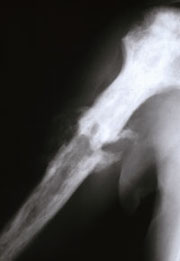
Plain radiograph shows a diaphyseal, ill defined, destructive, radiolucent lesion with permeative
margins. The lesion shows multifocal cortical disruption.
|
|
|
|
Pathological Findings: :
|
|
|

|
-
Low power view shows a highly cellular spindle cell neoplasm with focal
storiform cellular arrangement and lack of identifiable matrix (osteoid or chondroid).
|
|
|

|
-
Some areas were less cellular but had many bizarre cells.
|
|
|

|
-
On multiple sections, mitoses were frequent and included atypical forms.
Special stains revealed strong immunoreactivity with vimentin and faint, focal immunostaining with cytokeratins
AE1/AE3, Cam5.2.
|
|
|

|
-
Pre-existent bone shows features of Paget's disease - thickened bone trabeculae with irregular,
scalloped edges and mosaic lines of mineralization. Focal osteoclastic and osteoblastic activity was present.
|
|
|
Diagnosis: Paget's Sarcoma, Malignant Fibrous Histiocytoma type
|
|
Salient Points::
|
-
Paget's Sarcoma. High-grade sarcoma is a relatively rare complication
of Paget's disease occurring in 1% to 10% of cases. The incidence depends on the extent and severity of
Paget's changes (Dorfman HD, Czerniak B. Bone Tumors. 1998). Most patients are between 55 and 75 years of age.
Osteosarcoma is the most common type of Paget's sarcoma followed by malignant fibrous histiocytoma
(MFH), fibrosarcoma, chondrosarcoma, and giant-cell sarcoma. Skeletal distribution of these tumors mirrors
that of Paget's disease - axial skeleton, cranio-facial bones, and the major long bones (femur, tibia, and humerus).
Prognosis is generally poor due to the high grade of the tumors, difficulties at their surgical removal,
and the old age of patients.
-
MFH of bone can be generally subdivided into primary or secondary. Studies show that approximately 28% of
MFH are secondary lesions arising in a background of Paget's disease, radiation osteitis or bone infarcts
(McCarthy EF, Frassica FJ. Pathology of Bone and Joint Disorders. 1998). This type of sarcoma is also
found in dedifferentiated areas of low-grade neoplasms. High incidence of secondary MFH mandates a
careful search for a pre-existing osseous lesion.
-
MFH has a wide age distribution (ages 15 to 85) with a peak incidence in patients older than 40 years.
Although any bone may be involved, the tumor is most commonly found in the knee area (30% of cases),
proximal femur, humerus and pelvis.
-
The histologic hallmark of MFH is the mixture of spindle cells in storiform arrangement and pleomorphic
cellular areas. However, a variety of histologic patterns may be encountered. Those include myxoid, organoid,
and hemangiopericytoma-like patterns to name just a few. Mitoses are usually numerous, and atypical forms
are easily found. Eosinophilic, collagenous extracellular matrix may be present and may be confused with osteoid.
However, remember that the only absolute diagnostic feature of osteoid is mineralization. Special studies are of little
use in the diagnosis of MFH. Immunostains are used mainly to rule out other types of high-grade sarcomas.
-
Clinical Behavior. MFH is a very aggressive sarcoma with a high rate of metastases, usually to the lungs.
Studies show that secondary MFH behave in a more aggressive manner than primary MFH
(Dorfman HD, Czerniak B. Bone Tumors. 1998).
Available publications for the topic:
Paget's disease of bone, malignant transformation
Available publications for the topic:
MFH of bone
|
|
Selected References::
|
-
Wick MR, Siegal GP, Unni KK, et al: Sarcomas of bone complicating osteitis deformans
(Paget's disease): Fifty years of experience. Am J Surg Pathol 5:47, 1981
-
Haibach H, Farrell C, Dittrich FJ: Neoplasms arising in Paget's disease of bone:
A study of 82 cases. Am J Clin Pathol 83:594, 1985
|
|
|

