| The patient underwent
diagnostic enucleation OD on August 6, 2001 at University
Hospital, Newark (PDL). Pathological specimens were sent to
the Department of Pathology at UMDNJ (NM) as well as to the
Department of Pathology at the University of Illinois at Chicago
(RF). |
|
PATHOLOGICAL
EXAMINATION AT UMDNJ: The specimen received
at UMDNJ was found to contain an additional intraocular
glass foreign body embedded in inferotemporal subretinal
hemorrhage. The retina was totally detached. A single retinal
granuloma was noted without eosinophils in significant numbers
or other changes such as Dalen-Fuchs nodules. It was not
clear that the granuloma was a sign of sympathetic ophthalmia
versus a reaction to the intraocular silicone oil or blood.
PATHOLOGICAL
EXAMINATION AT UNIVERSITY OF ILLINOIS: The
choroid was sectioned en face at the University of Illinois.
Two choroidal granulomata were identified. They were found
to be loosely formed.
|
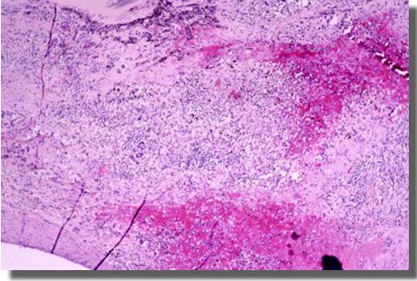
| CHOROIDAL
GRANULOMATA |
 |
 |
Focal
area of infiltration of lymphocytes, giant cells, and blood
within
the choroid (H&E 40x). |
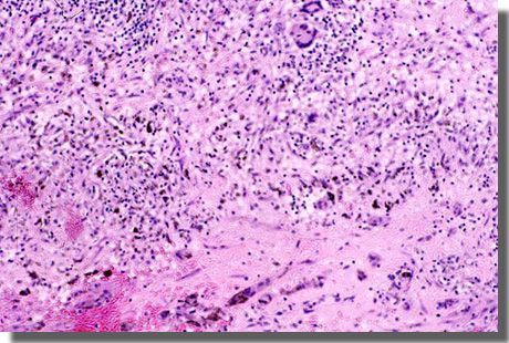
Ill-defined
non-caseating granulomata in the choroid (H&E 100x). |
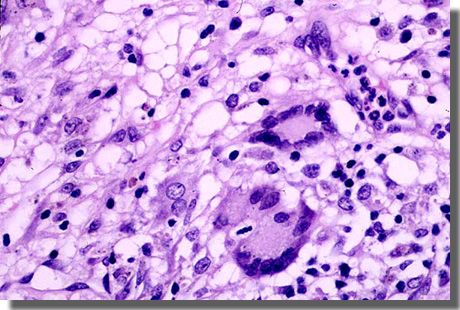 |
| Granuloma
with epitheliod histiocytes, giant cells and lymphocytes (H&E
400x). |
| MAINTENANCE THERAPY:
The pathological findings were felt to be consistent with
sympathetic ophthalmia. For this reason, the patient was maintained
on prednisone 60 mg po daily with Pepcid® 20 mg po, bid.
|
|
|