|
|
|
| FROM OUR Ophthalmic Imaging Center | |
|
Questions:
eyereplies@aol.com
|
|
| PHOTO TIP: In monochromatic photography, use a green filter and a red filter to visualize structures in the retina and choroid. |
|
|
|
| FROM OUR Ophthalmic Imaging Center | |
|
Questions:
eyereplies@aol.com
|
|
| PHOTO TIP: In monochromatic photography, use a green filter and a red filter to visualize structures in the retina and choroid. |
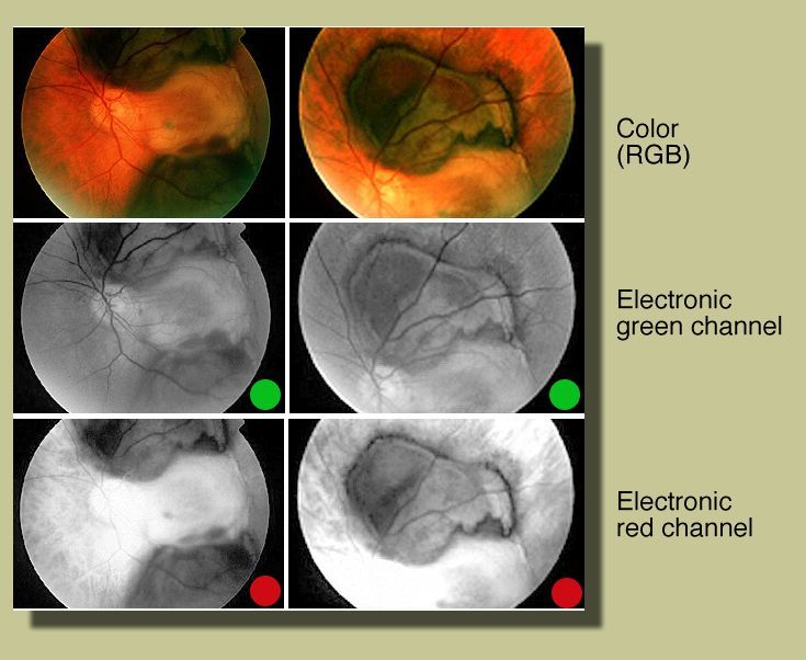
| Green and red electronic filters* | Monochromatic filters (RGB) |
 |
 |
|
Color fundus photos OS reveal a greenish subretinal mass. Green filter (electronic channel) highlights the subretinal
mass, retinal vessels, and retinal layers.
|
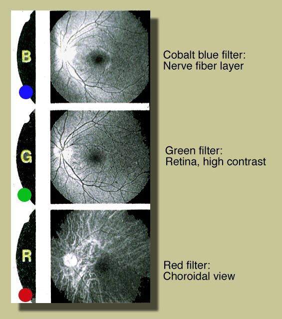
Typically, a cobalt blue filter demonstrates best the nerve fiber layers. The green filter—often called red-free filter—offers good contrast between the retinal layers, retinal vessels, and optic nerve head. The red filter allows documentation of the choroid and its vessels, usually seen as white vessels. | |
| Red filter (electronic channel) reveals choroidal details; subretinal mass remains visible (subretinal blood blocks red light from reaching the choroid). |
| * In Photoshop, press F2 and F3; alternatively, press Command 2 and Command 3 (Macintosh) |
| Please click here to return to case presentation |