|
 The
Ophthalmic Diagnostic Imaging Center encompasses
the Ophthalmic Imaging Division, the Telemedicine Project, the
Electrophysiology Service, the Orthoptics
Service, the Visual Field Service, and outpatient Minor Surgery.
The Ophthalmic Imaging Division is currently under the direction
of Beth Malpica. Its scope includes film and digital
contrast dye studies, fundus and external photography, scanning
laser ophthalmoscopy, confocal laser nerve fiber layer analysis,
corneal topography, ophthalmic ultrasound, and the Telemedicine
Project. This division provides services to physicians in the
Institute as well as throughout the State of New Jersey. The
Ophthalmic Diagnostic Imaging Center encompasses
the Ophthalmic Imaging Division, the Telemedicine Project, the
Electrophysiology Service, the Orthoptics
Service, the Visual Field Service, and outpatient Minor Surgery.
The Ophthalmic Imaging Division is currently under the direction
of Beth Malpica. Its scope includes film and digital
contrast dye studies, fundus and external photography, scanning
laser ophthalmoscopy, confocal laser nerve fiber layer analysis,
corneal topography, ophthalmic ultrasound, and the Telemedicine
Project. This division provides services to physicians in the
Institute as well as throughout the State of New Jersey.
Ocular assessments are accomplished with multiple imaging modalities:
the topographic scanning system (TopSS)
for evaluating the optic nerve in glaucoma, now with capabilities
for circulation studies using indocyanine green (ICG) for evaluating
the macula; the ImageNet Fundus
Imager, for
documenting retinal and choroidal blood flow with ICG and fluorescein
dyes; and the scanning laser ophthalmoscope (SLO)
for documenting flow circulation with ICG/fluorescein dyes and
for microperimetry. The SLO is an essential component of our RPE
transplantation program and is further used to train low vision
patients to fixate eccentrically. The Imaging
Division also features ocular echography as well as ultrasound
biomicroscopy (UBM)
and optical coherence tomography (OCT)
for specialized imaging of the anterior and posterior segments,
respectively.
Our imaging center maintains ancillary equipment for data enhancement,
with capabilities for scanning, editing, and enhancing videos,
color film, negatives, and computer-generated images as well as
for converting these modalities into any type of medium needed
for research, poster presentations, lectures, training, or review
purposes.
|
 |
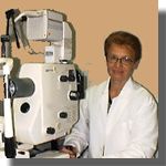
| Digital retinal camera with 20/35/50-degree field. Color and monochrome capabilities for
fluorescein angiography (FA) studies. |
 |
| |
| |
|
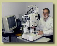 |
Film and ImageNet
digital retinal camera with variable field angle. Color,
monochrome, FA and ICG imaging capabilities. |
| |
| |
|
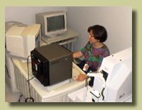
| SLO
with nonmydryatic capabilities for retinal field analysis
(visumetry and scotometry), FA and ICG studies. |
 |
| |
| |
|
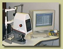 |
TopSS
for imaging the optic nerve head and producing 3-D animated
images for glaucoma studies. Also useful for nerve fiber layer
and ICG studies. |
|