| UNIVERSITY OPHTHALMOLOGY CONSULTANTS |
|
CASE OF THE MONTH CASE #2 |
| SUBRETINAL HEMORRHAGE | CHOROIDAL MELANOMA |
 |
||
| DIFFERENTIAL DIAGNOSIS | ||
|
This case illustrates subretinal hemorrhage mimicking choroidal melanoma. |
||
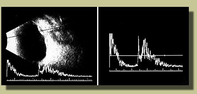
| The key is to see drusen in the fellow eye, choroidal neovascular membrane (CNV) in the study eye, a fluorescein angiogram that is typical of CNV in age-related macular degeneration (AMD), and an ultrasound that is typical of subretinal hemorrhage. |
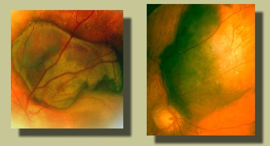
| Drusen in the fellow eye (OD) |
CNV
in the study eye (OS)
|
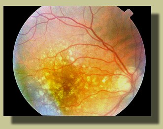
Fluorescein
angiogram (OS)
|
|
 |
 |
 |
The blockage of underlying choroidal fluorescence on the fluorescein angiogram is due to subretinal blood. |
|
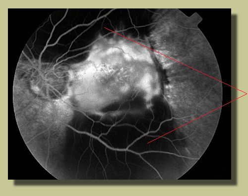
Echogram
(OS)
|
|||
 |
| Click to return to case presentation | |
| Please send comments to: Dr. Marco Zarbin at zarbin@umdnj.edu |
| Suggested Reading: |
| Hochman MA, Seery CM, and Zarbin MA. Pathophysiology and management of subretinal hemorrhage. Surv Ophthalmol 1997; 42(3):195-213. |