| External Eye
Examination |
|
Mild eyelid
swelling OU
Moderate conjunctival injection, more severe OS than OD |
| |
|
|
Visual Acuity |
OD:
20/200; Pinhole: 20/50
OS: Counting fingers; Pinhole: no improvement |
| |
|
| Intraocular
pressure with applanation tonometry |
OU:
8 mm Hg |
| |
|
| Slit
Lamp Examination |
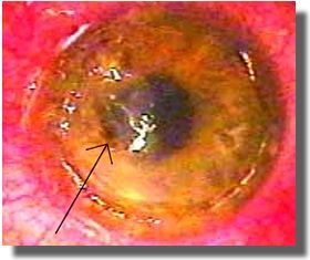
OD
|
| |
 |
| |
• Corneal flap in place
• Mild corneal rubeosis extending 360 degrees
• 5x4-mm infiltrate (arrow) in the flap interface at
the 3-
o’clock position with a 0.5-mm overlying epithelial
defect
• Deep anterior capsule with trace cells and flare
• Clear lens and anterior vitreous
|
| |
 |
| |
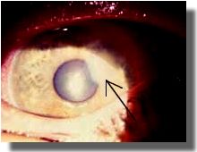
OS |
| |
 |
| |
•
Marked conjunctival injection
• Freely mobile, thickened corneal flap
• 5x5-mm area of flap necrosis (arrow) extending from
the 6-o’clock to the 10-o’clock position
• Diffuse infiltrate
• Limbal rubeosis
• Clear lens and anterior vitreous |
| |
|