| UNIVERSITY OPHTHALMOLOGY CONSULTANTS |
|
AGE-RELATED MACULAR DEGENERATION UPDATE: A REVIEW OF PATHOGENESIS, CLINICAL FINDINGS, AND TREATMENT
Supported in part by Research to Prevent Blindness, Inc. and the New Jersey Lions Eye Research Foundation. PAGE 5 |
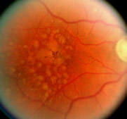
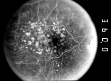
Figure 1. Fundus photograph of a patient with nonexudative age-related macular degeneration. A. The pale round subretinal spots, termed drusen, are a typical feature of the disease and are accumulations of cellular debris between the retinal pigment epithelium (RPE) and the subjacent choroid. Small hard and larger soft drusen are present. B. Fluorescein angiography demonstrates that many of the drusen are hyperfluorescent and more clearly demonstrates the presence of small drusen. Hyperfluorescence arises partly because of RPE rarefaction (with loss of pigment) over the drusen and partly because of dye leakage into the subRPE deposits. Drusen clusters are readily apparent on the fluorescent angiogram. |
|||||||||
|
age-related macular degeneration? Arch Ophthalmol 1992; 110:15-6. Lopez PF, Sippy BD, Lambert HM, Thach AB, Hinton DR. Transdifferentiated retinal pigment epithelial cells are immunoreactive for vascular endothelial growth factor in surgically excised age-related macular degeneration-related choroidal neovascular membranes. Invest Ophthalmol Vis Sci 1996; 37:855-68. Macular Photocoagulation Study Group. Risk factors for choroidal neovascularization in the second eye of patients with juxtafoveal or subfoveal choroidal neovascularization secondary to age-related macular degeneration. Arch Ophthalmol 1997; 115:741-7. Macular Photocoagulation Study Group. Visual outcome after laser photocoagulation for subfoveal choroidal neovascularization secondary to age-related macular degeneration: the influence of initial lesion size and initial visual acuity. Arch Ophthalmol 1194; 112:480-8. Meyers SM, Greene T, Gutman FA. A twin study of age-related macular degeneration. Am J Ophthalmol 1995; 120:759-66. Moore DJ, Hussain AA, Marshall J. Age-related variation in the hydraulic conductivity of Bruch's membrane. Invest Ophthalmol Vis Sci 1995; 36:1290-7 Nasir M, Sugino I, Zarbin MA. Decreased choriocapillaris perfusion following surgical excision of choroidal neovascular membranes in age-related macular degeneration. Br J Ophthalmol 1997; 81:481-9. Pauleikhoff D, Sheraidah G, Marshall J, et al. Biochemical and histochemical analysis of age-related lipid deposits in Bruch's membrane. Ophthalmology 1994; 91:730-4. Pauleikhoff D, Zuels S, Sheraidah GS, Marshall J, Wessing A, Bird AC. Correlation between biochemical composition and fluorescein binding of deposits in Bruch's membrane. Ophthalmology 1992; 99:1548-53. |
Reddy VM, Zamora RL, Kaplan HJ. Distribution of growth factors in
subfoveal neovascular membranes in age-related macular degeneration
and presumed ocular histoplasmosis syndrome. Am J Ophthalmol 1995;
120:291-301.
Sarks JP, Sarks SH, Killingsworth MC. Evolution of soft drusen in age-related macular degeneration. Eye 1994; 8:269-83. Sarks JP, Sarks SH, Killingsworth MC. Morphology of early choroidal neovascularization in age-related macular degeneration: correlation with activity. Eye 1997; 11:515-22. Sarks, JP, Sarks SH, Killingsworth MC. Evolution of geographic atrophy of the retinal pigment epithelium. Eye 1988; 2: 552-77. Seddon JM, Ajani UA, Mitchell BD. Familial aggregation of age-related maculopathy. Am J Ophthalmol 1997; 123:199-206. Seddon, JM, Ajani, UA, Sperduto, RD, Hiller, R, Blair, N, Burton, TC, Farber, MD, Gragoudas, ES, Haller, J, Miller, DT, Yanuzzi, LA, Willet, W. Dietary carotenoids, vitamins A, C, and E, and advanced age-related macular degeneration. JAMA 1994; 272:1413-20. The Choroidal Neovascularization Prevention Trial Research Group. Laser treatment in eyes with large drusen. Ophthalmology 1998; 105: 11-23. Weiter JJ, Delori F, Dorey CK. Central sparing in annular macular degeneration. Am J Ophthalmol 1988; 106:286-92. Young, RW. Sunlight and age-related eye disease. J Natl Med Assoc 1992; 84:353-8. Zarbin MA. Age-related macular degeneration: review of pathogenesis. Eur J Ophthalmol 1998; 8:199-205.
|
|||||||||

| PAGE 5 | Back to page 1 |
| residency program /// patient care services /// research |
| ophthalmic medical assistant program /// continuing medical education |
| facilities /// faculty /// library |